Phymatous Rosacea
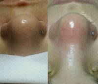
Phymatous rosacea: skin thickening, increased nose and surrounding tissues. The most common subtype is called rhinophyma,which affects the sebaceous glands and outer side of the nose and causes an increase in the nose and has a reddish or yellowish waxy appearance. Rhinophyma is more common in men.
Phymatous rosacea is a benign the tumorous formation, characterized by overgrowth and hypertrophy of the nose skin and all of its elements.
This nose is also called the “nose of drunkards”, “winy”, “pineal” nose. The disease is more common in men older than 40 years. Men suffer more often than women (about 6 times).
The etiology of the disease is not fully understood. The cause may be chronic inflammation of the skin, red spots, demodicosis, rosacea. Other factors predisposing to the disease, are stress, vitamin deficiencies, hormonal disorders, gastrointestinal disease, and – adverse environmental conditions, frequent exposure to cold, high humidity and dust.
The exact causes of phymatous rosacea are not installed. But we know that long-existing acne or rosacea, demodicosis often lead to phymatous rosacea.
Other predisposing factors of the disease are gastrointestinal, autoimmune diseases, alcoholism, hormonal imbalances, a sharp change of temperature conditions, hypothermia, exposure to harmful chemicals, chronic stress.
Against the background phymatous rosacea can occur concomitant inflammatory diseases – conjunctivitis, blepharitis. Direct connection of the disease has not been established with alcohol abuse.
Diagnosis and differential diagnosis Phymatous rosacea.

Phymatous Rosacea Pictures
The dermatologist usually diagnosticate “Rhinophyma” on the basis of visual inspection, the history of development disease and the patient’s history. Pressing the is allocated white pasty secret. The results of cytological investigation allow accurate make a diagnosis.
At microscopy are discovered epithelial cells in large amounts, sebum, saprophytic microflora, pathogenic microflora can not always be present, the presence of mites Demodex also observed not always.
Despite the vivid and specific clinical manifestations phymatous rosacea, it is necessary to differentiate with demodex as Phymatous rosacea,can be complicated by demodicosis, but the latter is not a major pathogenetic mechanism; cutaneous T-cell lymphoma, sarcoidosis and lymphatic leukemia can also occur growths of the skin tissue of the nose.
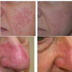
Phymatous Rosacea Cheeks
Additional tests, such as a tissue biopsy, with subsequent histological analysis helps to exclude or confirm the diagnosis of “Rhinophyma”.
Etiology and pathogenesis.
Rhinophyma develops slowly. Are possible alternation of rapid growth stages with periods of remission. Several years later, the process may stop, but changes do not disappear. The histological (tissue) study found that the increase of skin cells, particularly sebaceous glands, develops on the background of chronic inflammatory process.
Phymatous Rosacea Natural Treatment
There are four types of Phymatous rosacea: 1 (erythematotelangiectatic) rosacea, subtype 2 (papulopustular) rosacea, subtype 3 (phymatous) rosacea and subtype 4 (ocular) rosacea.
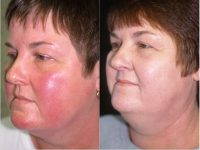
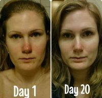
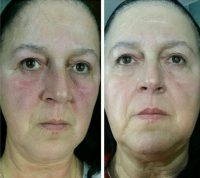
Phymatous Rosacea form appears bumpy or botryoidal soft formations, bluish-red color, with a shiny sebaceous surface and a large number the expanded vessels and sebaceous glands, which is allocated sebum under pressure. The skin overgrowth sometimes reaches enormous proportions, hanging down, closing his nostrils and mouth, making it difficult breathing, eating and disfigures the face.
Ocular rosacea form of rhinophyma struck the nose skin is smooth, hypertrophied tissue, and the skin is sealed due the tissue hypertrophy. Color – red-violet because of proliferation of blood vessels.

Phymatous Rosacea Nose Treatment
Erythematotelangiectatic form is characterized by copper-red or purplish-red color of varying intensity nose. The size of the nose is increased on the surface of large ectatic veins are visible. Unlike fibrous stage you can see a large number of superficial and deeper pustules. Patients complain of paresthesia, itching and soreness.
Papulopustular rosacea is characterized by an even moderate increase in the nose with protruding knobby accumulations of elastic tissue, which gradually becomes brownish-bluish color.
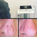
Treatment only operative with various alternatives for the removal phymatous rosacea. The operation is performed under local anesthesia. Removing Phymatous Rosacea begins with a invariably of healthy skin.
Phymatous Rosacea Early Symptoms
The surgeon forms a the proper shape of the nose, its outline, cutting off the hypertrophied tissue that does not contain lining glands which create spontaneous growth of the epithelium.
The vastness of the process requires in most cases remove damaged skin on the entire surface of the nose. The main methods of forming the nose contour, with a scalpel, laser excision, cryosurgery, dermabrasion, ultrasound destruction, electrocoagulation, radiosurgery.
The best result gives the radiowave surgery is a non-invasive way to remove and simultaneous coagulation tissue. With it became possible to remove the tissue very carefully, with an excellent cosmetic result.
Phymatous Rosacea Early Signs
The mechanism of this effect is due to the fact that during the passage of radio waves through the tissue, there is a high frequency power generation, which leads to the actual evaporation of cells.
Because the radiowave scalpel is not heated because thermal damage to tissue is absent. Accuracy and dosing of the radiowave method lets you delete only those cell masses neoplasm, which must be removed, the surrounding tissue is practically not affected, blood vessels are coagulated immediately, which negates the potential blood loss.
- Tissue is not carbonized and epithelization (healing) occurs twice as fast.
- Pain as during surgery and in the postoperative period – is absent.
Early Phymatous Rosacea Photos
Healing is performed by open method, without bandages. The wound surface is treated with ointments, accelerate the regeneration of the skin. Epithelization lasts 3-5 days, and full epithelization – 2-3 weeks. The surface of the skin of the nose formed smooth without scarring. In general, the healing process is fast enough with a good aesthetic result.
Prevention phymatous rosacea
Specific prevention phymatous rosacea is not, but timely treatment of rosacea reduce the risk of phymatous rosacea. After surgical treatment of rhinophyma the patients should avoid extreme temperatures, to reconsider the diet and, if required, to change jobs.
-

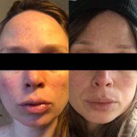
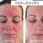
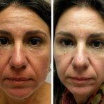
- Phymatous Rosacea Before And After
-
- Phymatous Rosacea Female
-

- Phymatous Rosacea Laser Treatment
-
- Phymatous Rosacea Nose
-

- Phymatous Rosacea Surgery
-
- Phymatous Rosacea Symptoms
-
- Phymatous Rosacea Treatment
-

- What Is Phymatous Rosacea Photo