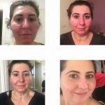
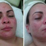
Rhinophyma Before And After
Rhinophyma is a benign tumor the skin of the nose, which is manifested by hypertrophy of all elements of the nose, it disfigures the face of a man.
Since all the elements of the skin increases, the size and width of the sebaceous glands are also hypertrophied. When rhinophyma is observed enhanced excretion of fat, the fat accumulate in the ducts and decomposes, resulting in an unpleasant smell arises.
For diagnosis “rhinophyma” is usually quite simple the dermatological inspection. Clarify diagnosis allows cytological examination.
The causes and mechanisms of development of rhinophyma
Rhinophyma is infiltrative-productive complication of rosacea and often diagnosed in men over the age of forty years. If in the patient’s anamnesis is not rosacea or acne rosacea, then rhinophyma is treated as a separate disease.
The etiology of the disease is not fully understood, but in people who are prone to frequent overheating and overcooling, temperature fluctuations such pathology is diagnosed more often; high humidity or excessive dryness and dustiness of air also increases the risk of rhinophyma.
In patients of rhinophyma in a history are usually present endocrine disorders, hormonal disorders, gastrointestinal disease.
Hypovitaminosis, stress and improper diet by themselves are not a major cause of rhinophyma, but in conjunction with the above factors increase the likelihood of its occurrence.

Bulbous Nose Rhinophyma Rosacea
There is no direct link between rhinophyma and chronic alcoholism, but given the fact that in alcoholism are joining various disease of organs and systems, excessive alcohol consumption and rhinophyma are closely linked.
Congenital vascular skin changes and transformation of congenital vascular nevus in most patients lead to changes the skin of face.
Clinical manifestations of rhinophyma
Externally rhinophyma looks like excrescence, it is due to hypertrophy and hyperplasia of the skin of the nose. Excrescence can be a single or in the form of nodes, then is diagnosed nodosum rhinophyma. Color of hypertrophied nodes can vary from red to dark brown and purple shades due to the expanded vessels.

Rhinophyma Treatment Surgery
Large follicles and sebaceous glands are corked with keratin, a region which struck by rhinophyma is a lymphatic interfollicular inflammatory infiltrate. If rhinophyma progresses, the granulomas are observed and often giant sebaceous glands are emptied into normal sinus.
When rhinophyma can be clearly seen that a large part hypertrophied tissue is composed of the expanded vessels with thin walls. The skin affected rhinophyma more susceptible to the development of neoplasms.
There are several varieties of rhinophyma. Glandular type of-Rhinofima is diagnosed more often, skin overgrowth in this case looks like a rough nodes. Consistency nodes is soft, and the surface of the affected area rhinophyma is glossy sebaceous with a cyanotic tinge, and less frequent purple.

Rhinophyma Surgery Before And After
Fibrous type of Rhinofima is less common, in this form the skin layers are hypertrophied, but the skin is is dense, which preserves the the nose configuration. The surface is smooth and shiny from enlarged pores may allocation of skin fat which gets an unpleasant odor on contact with air. On palpation there is a pronounced hyperplasia of the sebaceous glands of different densities.
Fibroangiomatus type of-Rhinofima is clinically similar to the fibrous form of rhinophyma, but on palpation nodes is more elastic and soft. The surface of the nose has a deep red color due to the large number of telangiectasias. This form rhinophyma differentiated from the others the presence of a large number of deep and superficial pustules. The contents of pustules saniopurulent that in process evacuation dries up to a crust. Patients in addition to shortness of breath complain of itch, pain and parastezii.

Rhinophyma Removal
Actinic form of rhinophyma has a benign course, layers of the dermis are uniformly become thickened, the nose becomes cyanotic color, telangiectasia are localized mainly on the wings of the nose.
The pustules are absent in this form of rhinophyma, and the pores of the sebaceous glands expanded slightly, so that the sebum is allocated moderately without formation of crusts on the surface of the nose. The main pathogenetic component of the actinic rhinophyma is actinic elastosis.
The course rhinophyma is long, with a series stages of remission and stages of growth. As a rule, the active rhinophyma growth is observed in the first years of the disease, the growth may stop completely after a few years, but the reverse development of rhinophyma is not observed.

Rhinophyma Pics
Rhinophyma complicates nasal breathing and eating, despite the fact that pathological process does not strike the cartilage.
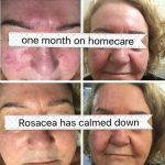
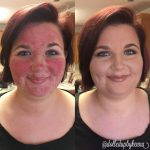
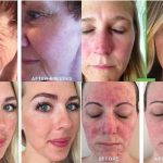
Treatment of rhinophyma
Therapeutic methods rhinophyma do not work, and therefore used different methods of surgical treatment. Dermabrasion the affected area of the skin gives good results at insignificant overgrowth tissue and in the initial period of the disease.
A subcutaneous excision of the overgrown tissue and a wedge excision of the affected parts with the imposition of intradermal sutures are shown at deep skin lesions of the nose and inability to use other methods of surgical treatment of rhinophyma.

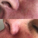
Rhinophyma Before And After Surgery Pictures
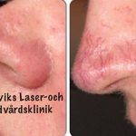
Laser therapy rhinophyma in the treatment process allows to model disfigured shape of the nose, achieved therapeutic and aesthetic effects.
Removing by laser overgrown dermis is shown when the insignificant part of the skin is struck. Epithelialization process occurs within 7-10 days after the procedure.
Radiowave method in the treatment of rhinophyma allows you to quickly and effectively eliminate all of the clinical manifestations and restore the shape of the nose.
When such treatment is almost no bleeding, no swelling, the probability of postoperative complications is minimal, and the recovery process is a short.
Rhinophyma Before And After
-

- Rhinophyma Of The Nose Pictures
-

- Bulbous Nose Rhinophyma Treatment
-

- Bulbous Nose Rhinophyma
-

- Images Of Rhinophyma
-

- Laser For Rhinophyma
-
- Rhinophyma (rosacea Of The Nose)
-

- Rhinophyma Before After
-

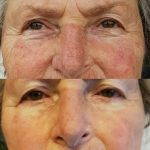
- Rhinophyma Before And After
-
- Rhinophyma Beginning Stages
-

- Rhinophyma Surgery
-
- Rhinophyma Laser Surgery
-
- Rhinophyma Laser Treatment
-

- Rhinophyma Nose
-

- Rhinophyma Of The Nose
-
- Rhinophyma Photos
-
- Rhinophyma Picture
-

- Rhinophyma Pictures And Symptoms
-

- Rhinophyma Pictures
-

- Rhinophyma Procedure
-

- Rhinophyma Reduction Surgery
-

- Rhinophyma Reduction
-
- Rhinophyma Rosacea Treatment
-

- Rhinophyma Rosacea
-

- Rhinophyma Surgery Photos
-

- Rhinophyma Symptoms
-
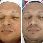
- Rhinophyma Treatment Before And After
-
- Rhinophyma Treatment Photos
-
- Rhinophyma Women Early Stages
-
- Rhinophyma Women
-

- Rhinophymatous Rosacea
-

- Rosacea And Rhinophyma
-

- Women With Rhinophyma